
Surgeons Just Used Live AI to Help Remove a Tumour Inside the Brain: Here's What Happened
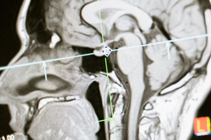
Surgeons at a London hospital have used artificial intelligence to analyse live video footage and guide them during brain tumour surgery for the first time.
The pioneering procedure took place at the National Hospital for Neurology and Neurosurgery in London. Doctors removed an 11-millimetre tumour from the patient's pituitary gland, at the base of his brain. The growth was close to the nerves controlling his vision and threatened his sight.
Rhys Hibbert, a 48-year-old father of two from Bedfordshire, discovered the growth after collapsing during an afternoon walk. The non-cancerous tumour grew on his pituitary gland, a marble-sized organ that regulates vital hormones. As the mass expanded, it pressed against his optic nerves, narrowing his vision and causing severe dizziness.
Without surgery, Hibbert faced irreversible sight loss.
'From a patient safety perspective, I can see absolutely the benefit,' Hibbert told the BBC. 'There are no road signs inside our head.'
He added: 'If patients are not prepared to join research how can doctors ever learn and how can medicine ever progress?'
Live Computer Vision Guides Surgeons Through Delicate Skull Base
Operating on the pituitary gland presents severe hazards for surgical teams. Doctors pass a camera-equipped endoscope through the nasal cavity to reach the skull base.
The target area sits millimetres away from the carotid arteries supplying blood to the brain. Conventional operations carry a 25 to 50 per cent chance of leaving tumour tissue behind, risking vascular injury. Pre-operative scans provide guidance, but living brain tissue shifts and bleeds during surgery.
To minimise dangers, University College London researchers developed a bespoke artificial intelligence system. The average UK surgeon performs only 10 to 20 of these operations annually.
The UCL software analysed endoscopic video feeds in real time, projecting coloured highlights over critical nerves and blood vessels on a secondary monitor. Crucially, the algorithm did not operate surgical instruments or make independent medical decisions.
'It is trained on more operations than most surgeons see or do in a lifetime,' said Professor Hani Marcus. 'It can act like an expert second pair of eyes.'
He performed the operation alongside neurosurgical resident Danyal Khan, noting that surgeons retained full authority throughout the procedure.
Patient Reports Restored Vision Following Landmark Hospital Procedure
The operation successfully cleared the tumour while leaving delicate surrounding structures completely undamaged. Hibbert reported rapid improvements in his eyesight and stamina following the intervention.
'When I opened my eyes after the operation it felt like I had got 360-degree vision,' Hibbert said. 'I hadn't seen that clearly for a year.'
He added: 'I also have my energy back and all of the side effects I was suffering before surgery have gone. It's given me my life back.'
Clinical Trials Required Before Widespread Operating Theatre Adoption
Despite the operation's success, researchers emphasise that a single case cannot validate routine clinical adoption. A 2026 review in Epidemiology and Health Data Insights highlighted that surgical AI requires rigorous prospective trials.
The review noted that high costs, algorithmic bias, and data variations currently limit hospital integration. Specialists agree that larger controlled studies must confirm whether real-time AI tools genuinely reduce complications.
The National Institute for Health and Care Research funded the procedure alongside Google, with expanded trials planned.