
16-Year-Old Boy Has a Tooth Growing Inside His Eyelid in Astonishing World First
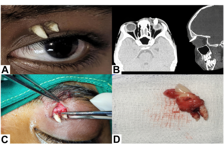
A 16-year-old boy in India has had a mature canine tooth removed from the upper part of his eye socket, a location where no such tooth is known to have been documented before. The case was published in the journal Ophthalmology.
The tooth sat in the space between his left eyelid and eyebrow, with two sharp projections showing. That spot lies in the superior orbit, or upper eye socket, which is why the case is believed to be a world first.
Teeth that erupt in abnormal locations are known as ectopic teeth. Earlier reports place them in the nasal cavity, nostrils, chin, palate and maxillary sinus.
A Surprising Diagnosis
The teenager first noticed painless swelling on his left upper eyelid that lasted a couple of months. About a month before he sought care, two sharp, whitish projections began protruding from the area.
The condition caused no pain, only cosmetic concerns. His parents worried about a possible tumour and whether it could affect his vision, according to case study co-author Meghana Tanwar, DO, DNB, of Aravind Eye Hospital in Madurai, India.
Doctors found the projections had a fairly smooth surface and felt hard to the touch, suggesting a tooth. They first considered a teratoma, a rare tumour that can contain bone and tooth tissue, or a foreign body in the eyelid.
A CT scan settled the question. 'The structure of a lone tooth in the superior orbit was quite obvious,' Tanwar said. Doctors later confirmed it was a mature canine tooth with no features suggesting a teratoma.
Why This Tooth Is Rare
Some ectopic teeth stay in the mouth: in about 2 to 6 per cent of people, the first permanent molars grow out of line in the expected row of teeth. This tooth, however, appeared completely outside the dental arch, which is far rarer.
To the best of the team's knowledge, it is 'the first and only ectopic tooth reported in the superior orbit', Tanwar said. Ectopic teeth have appeared in the eye socket before, but have typically been found in the bottom part known as the orbital floor.
'This was a first not just for us but the world over,' she said of the teenager's case. 'We were very surprised by it.'
The cause of the tooth remains unclear. A 2023 review listed several risk factors for ectopic teeth, including developmental disorders, trauma, infection, genetic factors and even developmental atavism, an evolutionary throwback.
Removal and Recovery
The tooth was removed under general anaesthesia after consultation with a maxillofacial surgeon. A meticulous dissection removed the entire tooth and its surrounding tissue without damaging normal functional tissue.
The procedure took about 20 minutes, and the wound had healed well at the two-week postoperative visit. The patient had a minimal scar and no problems over six months of follow-up.
The tooth was not harmful for the moment, as it was not pressing on other structures in the orbit. Had it migrated near muscles, nerves, vessels or the eye, it could have caused functional problems through mechanical compression.
Leaving an ectopic tooth in place can, depending on its location, damage nearby teeth or lead to cysts and sinus or nasal problems. Imaging showed no other ectopic teeth in the boy, so the likelihood of another is low.
Tanwar's team had seen a similar case before, removing a rare tumour-like growth called an odontogenic choristoma that contained a tooth in another patient's lower eye socket. A choristoma refers to normal tissue that appears in an abnormal location.