
Girl, 16, Hallucinated a Flying Green Dinosaur Before Tests Revealed Rare Brain Inflammation
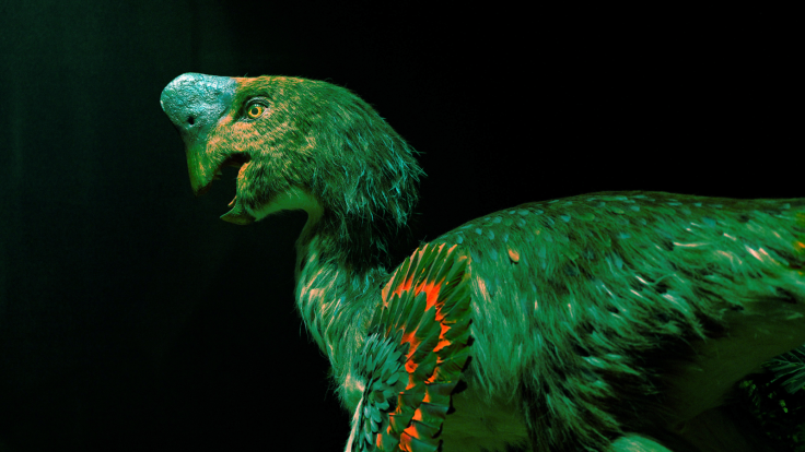
A 16-year-old girl who hallucinated a flying green dinosaur was diagnosed with a rare form of autoimmune encephalitis, according to a case report by emergency physicians at Loma Linda University Medical Center in California.
Her parents reported a sudden deterioration over the 48 hours before hospital admission, although the girl told clinicians that her hallucinations had begun in seventh grade. She has since recovered, though treatment continues.
A Dinosaur, Voices and a Sudden Decline
The teenager, an eleventh-grade student with no medical history, told clinicians she saw a flying green dinosaur, heard voices telling her to hurt herself and felt people were following her. She reacted to the creature during interviews as though it were present.
She told doctors the hallucinations began in seventh grade, but her parents reported no psychotic symptoms until 48 hours before presentation.
Her parents said her excellent grades had slipped over the previous month and that she had withdrawn from social activities. The family had travelled to the Philippines three weeks earlier, and several members, including the girl, developed a cough.
Because anti-NIF autoimmune encephalitis has been reported in adults with infections, including ehrlichiosis, doctors treated her empirically with doxycycline. However, the case report did not establish that an infection caused her condition.
Weeks of Tests Before Autoimmune Encephalitis Was Found
On arrival her heart rate stayed in the 140s and she was sweating and tremulous, which led staff to suspect a physical cause rather than a purely psychiatric one. A CT scan, an MRI, an electroencephalogram and initial spinal fluid tests were all unremarkable.
On the second day of admission she became completely catatonic and was given lorazepam, after which the catatonia improved over several days.
Psychiatrists considered psychosis but stressed the need to exclude an organic cause, and olanzapine was added on hospital day 11. A stool test on day four also detected cryptosporidium, which was treated.
Psychiatrists recommended transfer to an inpatient psychiatric unit on day eight, but difficulty feeding herself prevented it. On hospital day 18, a cerebrospinal fluid panel tested positive for anti-heavy chain neuronal intermediate filament antibodies, with a titre of 1:16.
Because the antibody is strongly associated with cancer, scans of her chest, abdomen and pelvis were carried out, but showed no sign of malignancy. Outpatient PET imaging later found no evidence of a neuroendocrine tumour or other cancer.
Treatment, Recovery and a Warning for Emergency Doctors
Over the following month she received intravenous steroids, immunoglobulin, rituximab and five sessions of plasmapheresis, and was discharged on hospital day 57. More than a year after admission, she reportedly remained free of symptoms and no longer took psychiatric medication, although immunotherapy continues.
The authors describe autoimmune encephalitis as uncommon, citing an estimated incidence of five to eight cases per 100,000 people. This estimate applies to autoimmune encephalitis generally, not specifically to the anti-NIF-associated subtype.
The authors stress that emergency doctors need not diagnose autoimmune encephalitis themselves, but should recognise features such as sudden onset, recent infection and abnormal vital signs, and direct further testing. The report appeared in Clinical Practice and Cases in Emergency Medicine, and the family consented to publication.